Among the most advanced research facilities in the nation, CBIS supports 11 state-of-the-art Research Core Facilities that are available to all RPI faculty, staff, and students, and to other academic, government, medical, and industry collaborators. These Core Facilities enable researchers to further innovation and discovery across disciplines in order to find solutions to pressing global challenges.
Our Core Facilities occupy 27,350 square feet within our 218,000-square-foot facility. Each Core Facility is led by a director holding a Ph.D. in their respective area and actively engaged in research. All Core Facilities are open to students, post-doctoral fellows, and principal investigators and operate on a fee-for-service basis. If you would like to become a user, the Core Directors will train you on the specific techniques required for your research.
Researchers can conduct their work at our Core Facilities in Troy, New York, or send in research samples.
Contact Information:
Marimar Lopez, Ph.D.
Director, Research Cores
biotechcores@rpi.edu
lopezm4@rpi.edu
Resources

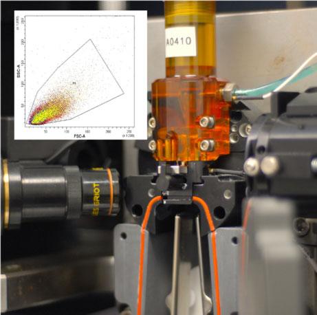


Rensselaer’s Stem Cell Research Core is a recharge facility designed to provide equipment and training and informal discussions to investigators, academic and industry, currently engaged in or seeking to pursue stem cell and mammalian cell biology