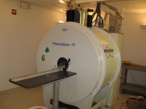
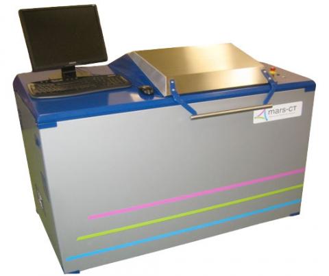
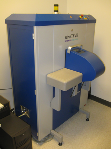
The Bioimaging Research Core has 14T and 7T magnetic resonance imaging (MRI) scanners, a micro computed tomography (μCT) scanner, and a highly novel spectral CT scanner with single photon counting capabilities (offering x-ray imaging at histology levels) enabling high-resolution and novel technologies for gaining contrast and sensitivity in imaging of live animals, excised tissues, and materials.
Support and fee-for-service options are available for experiment design, training in instrument operation, data acquisition, image processing/reconstruction, image analysis, and data management including the preparation of manuscripts and grant applications. The instruments are complemented with an animal procedure room, biometric monitoring equipment, inhaled anesthesia machines, and advanced computational resources including redundant files servers and multiple data workstations with a variety of software packages for processing and analysis.
Assistance and/or training in preparation of IACUC applications, live animal handling, anesthesia delivery, and biometric monitoring is available by trained technicians from the Bioresearch Core facility. Research animals are cared for in short-term and long-term transient housing rooms provided with highly sophisticated, barrier housing systems. All procedures are reviewed and require approval by the Rensselaer Institutional Animal Care and Use Committee (IACUC) and the attending veterinarian.
Additional Imaging Technologies available in the:
Contact Information:
Scott A. McCallum, Ph.D.
BioImaging & NMR Research Core Director
518-276-2856
mccals@rpi.edu
Access to the Facility and Equipment
Access to all equipment must be approved by the Core Director. Approved users will have their ID cards activated to access the instrument room.
Training
All users must receive proper training before being permitted access to the equipment. Training is provided by the core director.
Scheduling
Advance reservation using our online scheduling calendar is required. (If you do not have an account, contact the Core Director.) The billing is based on the length of occupation time, which is calculated based on the equipment computer log records. Cancellation at least 48 hours prior to the experiment is required. Users failing to do this will be charged (at the hourly rate) for the booked time.
Cleaning Up
Users are responsible for keeping the equipment and equipment areas safe and very clean. Failing to maintain this policy will result in loss of use privileges. Users are encouraged to remove their data from the hard disk of the computer immediately after each session. Long-term storage of large quantity of data on the equipment computer is not recommended as the data can be removed from the system without any warning.
Billing
Users will be required to provide billing information prior to requesting instrument time. Users with multiple billing accounts should specify the account number to be charged upon sign-up. Both internal and external users will be billed on a monthly basis. Providing fraudulent and/or inaccurate billing information may result in the loss of use privileges.
Technology