The mission of the Biomedical Imaging Center at Rensselaer Polytechnic Institute (RPI) is to define biomedical imaging frontiers, realize clinical/preclinical potentials, and train the next generation of imaging scientists and engineers. BIC occupies office and laboratory space of ~2,000sf in the Center for Biotechnology and Interdisciplinary Studies (CBIS), which ranks among the world’s most advanced research facilities focused on application of engineering, physical and information sciences to the life sciences.
Our laboratory is being setup to accommodate a medical CT system whose gantry was donated by GE Global Research Center, a high-performance, interior-scanning, general-purpose, and hybrid-modality (HIGH) mesoscopic imaging platform, and a test bed for nano-CT development.
resource2.png
X-ray & Hybrid Imaging (XHI) Laboratory:
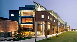
Biomedical Imaging Cluster within the CBIS building on the main campus of RPI.
The HIGH mesoscopic imaging platform has been uniquely designed for studies on living organisms or tissue samples of 1mm-1cm in size using spectral micro-CT, bioluminescence tomography (BLT), fluorescence molecular tomography (FMT), x-ray luminescence CT (XLCT), and x-ray fluorescence CT (XFCT).
resource3.png
HIGH platform
High-performance, interior, general, and hybrid (HIGH) mesoscopic imaging platform.
A micro-focus x-ray source (L10101, Hamamatsu Photonics, Inc.) is operated at 100kVp and 100-200μA. The focal spot size is 5-30μm.
Source & Detector: The -100ºC cooled EMCCD array (Andor, Belfast, Northern Ireland, U.K.) has 2048×2048 elements, 13.5×13.5μm2 per element for a 27.6×27.6mm2 active area, and a maximum frame rate of 4.7fps with 2×2 binning. Two EMCCD arrays are used for x-ray and optical signal detection, respectively. For x-ray imaging, an EMCCD array is optically connected to a scintillator screen made of GdO2S or CSI (TI). There is a thermoelectrically cooled cadmium telluride (CdTe) detector (X-123CdTe, Amptek Inc.). This detector module includes a preamplifier with pile-up rejection, a digital pulse processor, and a multichannel analyzer (MCA) (PX4, Amptek Inc.). Also, there is a MARS spectral x-ray camera acquired under a research agreement with MARS Bioimaging Ltd (http://marsbioimaging.com). The MARS camera is based on the new generation Medipix chip licensed out of CERN, Switzerland and the technology developed by the University of Canterbury (UC) and its partners. As the world’s leading spectral (multi-energy or true color) x-ray imaging technology, the MARS camera has six Medpix ASICS/CdTe detectors of 256×256 elements of 55×55μm2 per element for 14x14mm2 active area, and in 8 energy bins.
Stages: The distance between the source and the rotation center can be changed by a translation stage (IMS400, Newport, Inc.) for different resolution. The minimum focus-to-object distance is 6.8mm. The high-precision rotation stage (RV240HAT-F, Newport, Inc.) allows rotation of an object with an angular resolution of 0.0005º and repeatability error <0.0002º. The integrated high precision x-y translation stage (VP-25XA, Newport, Inc.) supports a resolution of 0.1μm and repeatability <0.2μm.
Lens & Filters: A set of lens are used for 0.5-40X magnification. These lenses are mounted on a turret. An additional macro-lens (Nikon normal 55mm f/1.2 AIS manual focus lens) provides a second chain for imaging. For multi-spectral optical imaging, various optical filters are needed in front of the CCD camera. These filters include a Green-Red dichroic plate beam splitter (Edmund Optics Inc.) and a Red-NIR dichroic plate beam splitter (Edmund Optics Inc.). The former reflects the signal of 530-595nm in wavelength, while the latter is for the signal of 595-664nm in wavelength.
Computer & Language: The HIGH platform is controlled by a PC workstation (HP8200, Hewlett-Packard, USA) in LABVIEW™ (National Instruments, USA).
Over the past three years, we have been upgrading Information Technology (IT) infrastructure to provide indexed repository for all results/files, standardize data storage/backup, consolidate all web contents, create user friendly interface for data sharing, and improve communication. We use Alfresco (http://www.alfresco.com), which is an open-source Enterprise Content Management System (CMS), WordPress (http://wordpress.org), and a commercial virtual video conference system ooVoo (http://oovoo.en.softonic.com). This feature is especially useful for interaction with our out-of-state and international collaborators, as shown below.
The Computational Center for Nanotechnology Innovations (CCNI) is a partnership between Rensselaer Polytechnic Institute, IBM, and New York State (https://ccni.rpi.edu/w), as shown in Figure R6. The three partners have created one of the world’s most powerful university-based supercomputers, based on the main campus at RPI and at its Rensselaer Technology Park. This center is an important resource for imaging researchers to perform most complicated numerical simulation and modeling in a highly efficient fashion.