Core Facilities

Among the most advanced research facilities in the nation, CBIS supports 11 state-of-the-art Research Core Facilities that are available to all Rensselaer Polytechnic Institute faculty, staff, and students, and to other academic, government, medical, and industry collaborators. These Core Facilities enable researchers to further innovation and discovery across disciplines in order to find solutions to pressing global challenges.

Our Core Facilities occupy 27,350 square feet within our 218,000-square-foot facility. Each Core Facility is led by a director holding a Ph.D. in their respective area and actively engaged in research. All Core Facilities are open to students, post-doctoral fellows, and principal investigators and operate on a fee-for-service basis. If you would like to become a user, the Core Directors will train you on the specific techniques required for your research. 

Researchers can conduct their work at our Core Facilities in Troy, New York, or send in research samples.

Contact Information:

Marimar Lopez, Ph.D. 
Director, Research Cores 
biotechcores@rpi.edu 
lopezm4@rpi.edu

Resources
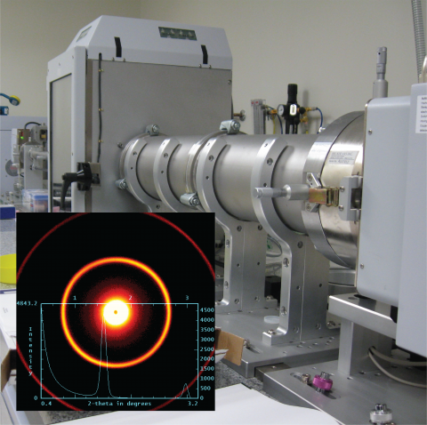
Nanostar with relevant data
The Analytical Biochemistry and Nano-Bio-technology Research Core has a variety of instruments for analyzing samples (biological and non-biological) at the atomic, molecular, and “nano” scales. Central to this core facility is a group of instruments that are typically considered fundamentally important for biophysical characterization of biological samples. These include: UV-visible absorbance, Circular Dichroism, Fluorescence (including lifetime and polarization), Infrared (FTIR), Analytical Ultracentrifuge, (SAXS), Isothermal Titration Calorimeter, Plate Reader for high-throughput UV-vis absorbance, fluorescence, and luminescence, HPLC, and FPLC-type chromatograph. Characterization of materials and their analysis are performed with Thermo-gravimetric Analyzer (TGA), Differential-Scanning Calorimeter (DSC), Wide-angle X-ray diffraction (XRD) and SAXS, Dynamic light scattering (DLS), Infrared microscope (paired to the FTIR Spectrophotometer).
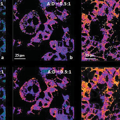
Examples of microscopic bio images
The Bioimaging Research Core has 14T and 7T magnetic resonance imaging (MRI) scanners, a micro computed tomography (μCT) scanner, and a highly novel spectral CT scanner with single photon counting capabilities
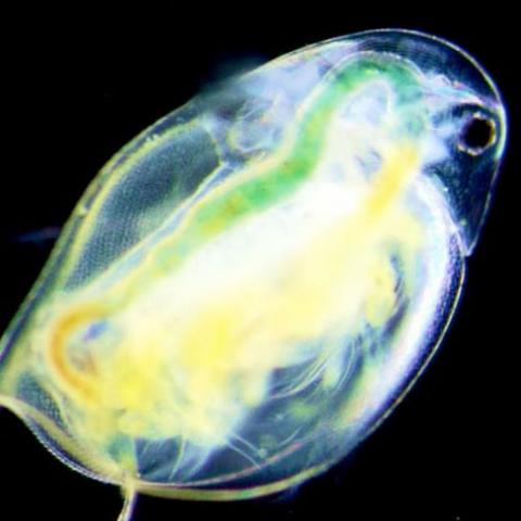
Bio image of plankton
The Bioresearch Core facilities include 11000 square feet of rodent research support space with barrier housing in ventilated rack systems, surgery and procedure suites, necropsy and anesthesia rooms, sterilization and washing facility, Hydropac (bottle-free) water system, an aquatic research complex, and quarantine facilities.
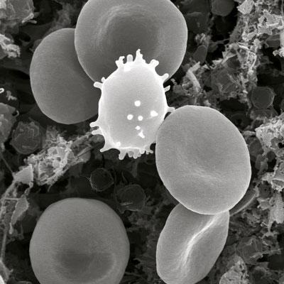
Red blood cells, white blood cells and platelets
The Cell and Molecular Biology Core Facility provides highly advanced molecular biology instruments to support experimental demands ranging from cell and tissue culture techniques to advanced cell transfection and genomics applications capabilities.
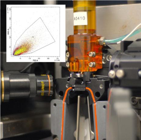
Flow cytometry equipment with an example of the results.
The Flow Cytometry Core offers researchers with powerful instruments to measure multiple physical characteristics of individual cells in suspension. The BD LSR II instrument provides multi color immune-phenotyping, protein expression level, cell cycle analysis, apoptosis studies, and measurements diverse cellular functions.
Image of a DNA Helix
The Genomics Research Core Facility provides users a full line of next generation sequencing (NGS) solutions from our state-of-the-art laboratory and computational infrastructure. We offer services from nucleic acid isolation, sample QC, library preparation, Illumina and Oxford Nanopore sequencing, data processing, to custom bioinformatics analysis. We also provide training and consultation sessions on bioinformatics, sequencing preparation, and continuous advancement in NGS.
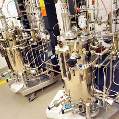
Fermentation equipment in the microbiology core
The Microbiology and Fermentation Core is a service facility dedicated to growing microorganisms and processing them for further analysis of cells and sub-cellular components such as proteins, carbohydrates, etc.
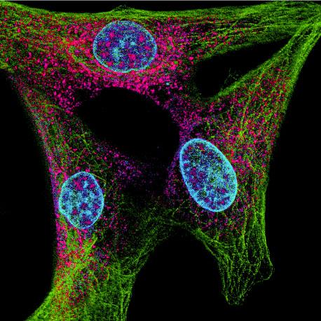
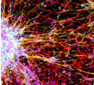
Example of the images produced by the microscopy equipment
The Microscopy core specializes in cross-disciplinary interaction through advanced imaging technology. The Core provides cellular and subcellular visualization up to super-resolution magnification in fluorescence, color brightfield, polarization and phase contrast channels. Advanced light microscopy with top of the line STED, Multi-Photon, Confocal and light sheet microscopes are accessorized with live cell incubators that maintain temperature, humidity, and CO2 to facilitate imaging of highly sensitive biological samples. In addition to light microscopy the Atomic Force Microscopy (AFM) is a technique the Core offers to measure surface topography and cell stiffness. AFM can be used to simultaneously perform optical transmission imaging of live cells. A dedicated image analysis workstation is available with deconvolution, particle tracking, colocalization, 3D reconstruction, and other analytic and image processing softwires.
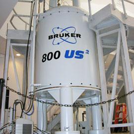
Bruker 800 MHz Spectrometer
The Nuclear Magnetic Resonance (NMR) Research Core provides RPI researchers in addition to academic and industrial users throughout the community access to state-of-the-art ultra-high field NMR instrumentation.
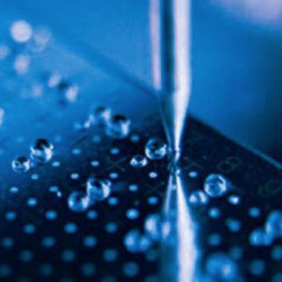
Bruker protein characterization
The Proteomics Research Core Facility offers proteomics analysis and mass spectrometry services to the scientific community. The Facility supports a variety of research projects in areas of manufacturing synthetic heparin, carbohydrate analysis and structural biology, osteoporosis, osteoarthritis and Alzheimer’s disease, drug discovery and delivery, material science, and environmental analyses. The facility houses several mass spectrometers, HPLC systems, and software packages for data search and protein identification.
Liquid-handling robot in the stem cell research laboratory at CBIS

The Stem Cell Research Facility Core is a recharge facility designed to provide equipment and training to all investigators, academic and industry, currently engaged in or seeking to pursue stem cell and mammalian cell biology research.
Required Acknowledgement and Authorship

Please acknowledge the CBIS Core Facilities in all publications and grant applications where our equipment and/or personnel have facilitated the work. These acknowledgements are very important because documenting our contributions helps to ensure that the resources of the Core Facilities are sustainable.

  • Equipment: If you used Core Facility equipment, please note this in the Materials and Methods. e.g., Thermogravimetric analysis was carried out using a TA Instruments TGA-Q50 (Rensselaer CBIS Analytical Biochemistry Core Facility).
  • Personnel: Please consider including CBIS personnel as co-authors on your publications when they have made a significant intellectual contribution to the research. Include CBIS Core Facility directors or staff as co-PI or co-investigators in grant applications when they provide a significant contribution to the grant proposal and scientific/intellectual leadership for the proposed work. Please follow these guidelines: ABRF Recommended Guidelines for Authorship on Manuscripts. Also, our Core Facility personnel always appreciate when they are mentioned in the Acknowledgements section of publications.
  • Required Funding Authorization Form: Rensselaer researchers must fill out the CBIS Cores Authorization Form (PDF) to use the CBIS Core Facilities.
Back to top